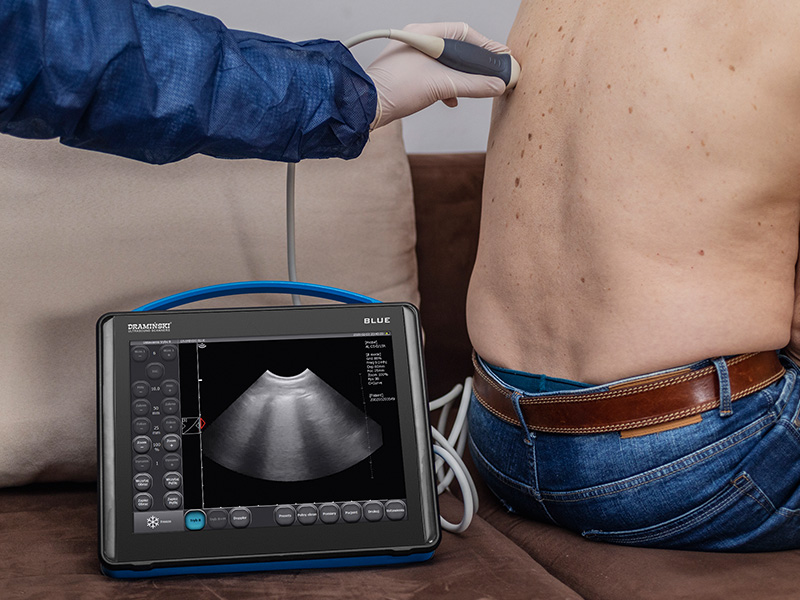
Ultrasound symptoms of the infection may be seen before the patient will develop others like cough or fever.
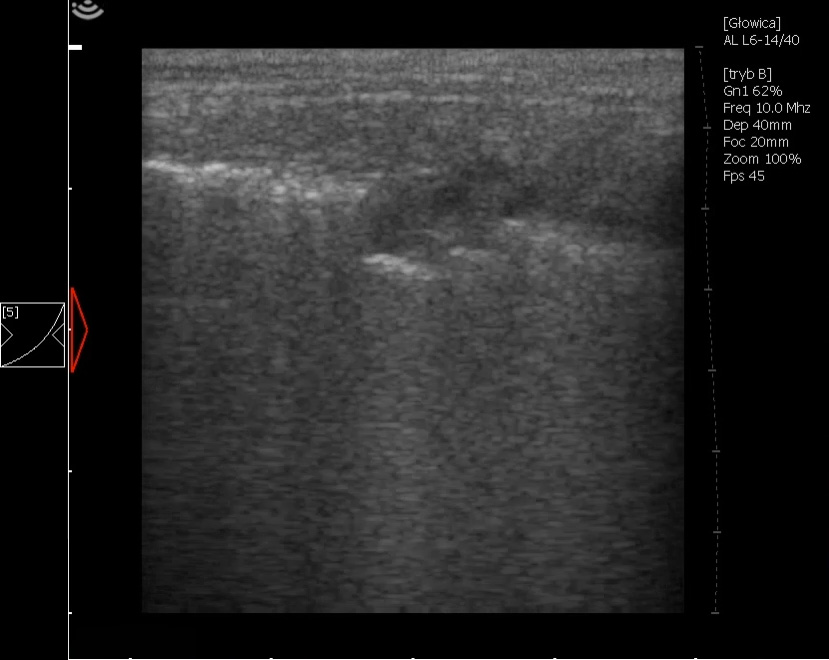
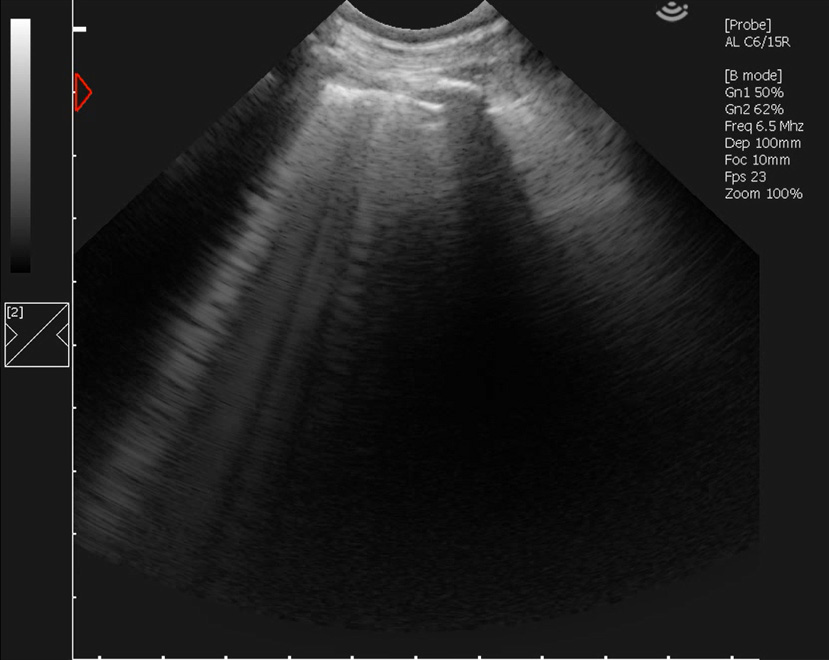
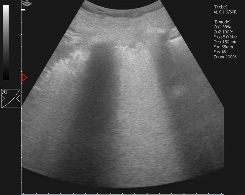
The most common ultrasound image is: B lines, subpleural consolidations (with or without air bronchogram), thickened pleura. B lines can be multifocal or confluent. Pleural effusion may be seen as well but in less cases. See the images!
The lesions are located mostly at posterior lower area (left and right), posterior upper area (left and right), axillary lower and upper area (mostly on the right side). Go beyond the protocols and scan as much as you can.
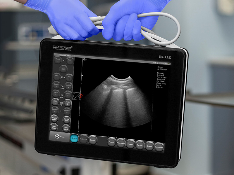
When monitoring your patient – in those who are reacting on treatment you will start to see A lines instead of B lines.
First publications were made on CT but many intensive care specialists underline that ultrasound imaging is quicker, easier to perform (as you do not have to transport your patient), cheaper and sensitive. Point-of-care ultrasound units give you an opportunity to examine patients ER, ICU and at their homes.
Use your ultrasound wisely.